
The head is one of the most complex and vital regions of the human body, housing critical structures responsible for sensory perception, communication, and cognitive function. It is composed of the skull, which protects the brain, and the face, which includes features essential for expression, breathing, and eating. The head also contains specialised sensory organs—such as the eyes, ears, nose, and mouth—that allow us to interact with and interpret the world around us. Additionally, it is supported by a network of muscles, nerves, and blood vessels that enable movement, sensation, and nourishment. Understanding the anatomy of the head provides insight into how we see, hear, smell, taste, speak, and think, making it a fascinating and essential area of study. Here is a basic overview of the makeup of the head.
1. Skull (Cranium)
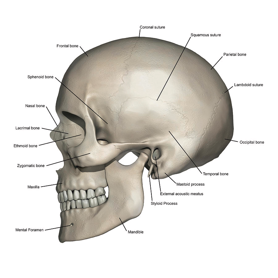
- Frontal Bone: Forehead and upper part of the eye sockets.
- Parietal Bones: Top and sides of the skull.
- Temporal Bones: Sides and base of the skull, housing the ear structures.
- Occipital Bone: Back and base of the skull, contains the foramen magnum (where the spinal cord connects to the brain).
- Sphenoid Bone: Base of the skull, behind the eyes.
- Ethmoid Bone: Between the eyes, contributes to the nasal cavity and orbits.
2. Face
- Orbits: Eye sockets, formed by multiple bones.
- Nasal Bones: Bridge of the nose.
- Zygomatic Bones: Cheekbones.
- Maxilla: Upper jaw, holds upper teeth.
- Mandible: Lower jaw, holds lower teeth.
- Temporomandibular Joint (TMJ): Joint connecting the mandible to the skull.
3. Muscles of the Head
- Occipitofrontalis: Raises eyebrows and wrinkles forehead.
- Orbicularis Oculi: Closes eyelids.
- Orbicularis Oris: Closes and protrudes lips (kissing muscle).
- Buccinator: Compresses cheeks (used in blowing and sucking).
- Masseter: Chewing muscle, elevates the mandible.
- Temporalis: Assists in chewing, elevates and retracts the mandible.
4. Sensory Organs
- Eyes: Vision, located in the orbits.
- Ears: Hearing and balance, divided into outer, middle, and inner ear.
- Nose: Smell and respiration, includes nasal cavity and sinuses.
- Mouth: Taste, speech, and digestion (teeth, tongue, salivary glands).
5. Nerves
- Cranial Nerves: 12 pairs of nerves originating from the brain, responsible for sensory and motor functions in the head and neck.
Examples: Olfactory (smell), Optic (vision), Facial (facial expressions), Trigeminal (facial sensation and chewing).
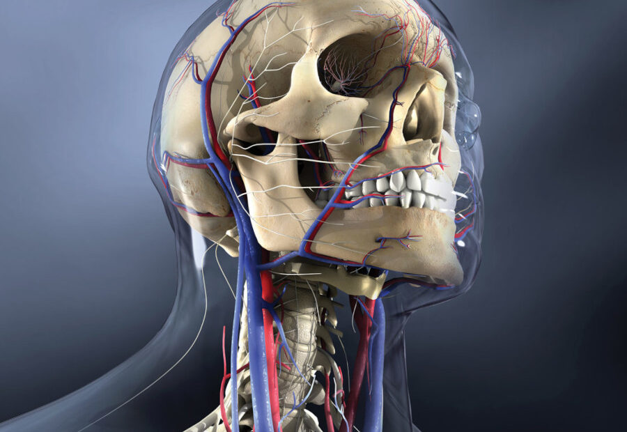
6. Blood Supply
- Carotid Arteries: Supply oxygenated blood to the head and neck.
- Vertebral Arteries: Supply blood to the posterior part of the brain.
- Jugular Veins: Drain deoxygenated blood from the head and neck.
7. Brain
- Cerebrum: Largest part, responsible for thought, memory, and voluntary actions.
- Cerebellum: Coordination and balance.
- Brainstem: Connects the brain to the spinal cord, controls vital functions like breathing and heart rate.
8. Other Structures
- Sinuses: Air-filled cavities in the skull (frontal, maxillary, ethmoid, sphenoid).
- Salivary Glands: Parotid, submandibular, and sublingual glands produce saliva.
- Teeth: Incisors, canines, premolars, and molars for chewing.
The head is a remarkable and intricate part of the human body, serving as the central hub for sensory input, communication, and higher cognitive functions. From the protective skull and expressive facial muscles to the complex sensory organs and the brain itself, every structure plays a vital role in our daily lives. Understanding the anatomy of the head not only highlights its importance but also underscores the interconnectedness of its systems. Whether it’s the way we see, hear, speak, or think, the head is a testament to the incredible complexity and efficiency of human biology. Continued study of this region remains essential for advancements in medicine, neuroscience, and overall human health.
References
– Gray’s Anatomy: Standring, S. (2020). Gray’s Anatomy: The Anatomical Basis of Clinical Practice (42nd ed.). Elsevier.
– Netter’s Atlas of Human Anatomy: Netter, F. H. (2019). Netter’s Atlas of Human Anatomy (7th ed.). Elsevier.
– Clinically Oriented Anatomy: Moore, K. L., Dalley, A. F., & Agur, A. M. R. (2018). Clinically Oriented Anatomy (8th ed.). Lippincott Williams & Wilkins.
– Human Physiology: Silverthorn, D. U. (2019). Human Physiology: An Integrated Approach (8th ed.). Pearson.
– Cranial Nerves Overview: Vilensky, J. A., Robertson, W. M., & Suarez-Quian, C. A. (2015). The Clinical Anatomy of the Cranial Nerves. Wiley-Blackwell.
This article is copyrighted by Ital is Vital, 2016 - 2026. Want to re-post this article? Visit our guidelines.
DISCLAIMER: THIS WEBSITE DOES NOT PROVIDE MEDICAL ADVICE
The information, including but not limited to, text, graphics, images and other material contained on this website are for informational purposes only. The purpose of this website is to promote broad consumer understanding and knowledge of various health topics. It is not intended to be a substitute for professional medical advice, diagnosis or treatment. Always seek the advice of your physician or other qualified health care provider with any questions you may have regarding a medical condition or treatment and before undertaking a new health care regimen, and never disregard professional medical advice or delay in seeking it because of something you have read on this website.